New AI Model Enhances Medical Image Diagnosis with Hierarchical Attention
Researchers have developed an advanced AI model, based on the Swin Transformer, to significantly improve the accuracy of medical image classification by focusing on diverse feature levels, from macro-scale lesions to minute anatomical details.

A new AI model significantly improves medical image classification by adeptly handling varied lesion scales and intricate tissue structures, promising more accurate and robust diagnostic support.
Advancing Medical Diagnostics with AI
The landscape of precision medicine and intelligent diagnostic tools is rapidly evolving, with automated medical image classification emerging as a crucial aid for clinical decision-making. However, the path to fully reliable AI in this domain is fraught with challenges. Medical images present a complex array of difficulties for deep learning models, including substantial variations in lesion sizes, intricate interdependencies between tissue structures, and the critical need to identify extremely subtle anatomical characteristics. These factors often limit the effectiveness of existing AI solutions, prompting researchers to seek more sophisticated approaches.
Traditional deep learning models, while powerful, often struggle with the multi-faceted nature of medical imagery. For instance, a model might excel at identifying large, obvious tumors but miss tiny, early-stage indicators of disease. Conversely, models designed for fine-grained detail might be overwhelmed by broader contextual information. Bridging this gap requires an architecture that can process information at multiple resolutions and conceptual levels simultaneously, ensuring that no critical piece of diagnostic information is overlooked.
Responding to these limitations, a team of researchers has proposed an innovative solution: a hierarchical complementary feature enhancement framework. This new architecture builds upon the foundation of the Swin Transformer, a powerful deep learning model known for its ability to process image data efficiently. The core idea is to equip the AI with an enhanced capability to model multi-scale lesions and engage in multi-level feature interactions, thereby providing a more comprehensive understanding of medical images than previously possible.
A Multi-Layered Approach to Feature Extraction
The proposed framework introduces a sophisticated method for collaborative feature learning, operating across three distinct representation levels. This tiered approach ensures that the model captures a complete picture, from broad overview to granular specifics. The first level, macro-scale lesion perception, focuses on identifying and understanding larger pathological features. This involves grasping the overall context and major anomalies within an image, much like a clinician's initial scan of a radiograph for prominent issues.
Following this, the model engages in global contextual interaction. This stage is crucial for understanding the relationships between different parts of an image, recognizing how various structures interact and influence each other. It allows the AI to develop a more holistic understanding, moving beyond isolated findings to see how they fit into the broader anatomical or pathological context. Finally, the third level, local detail refinement, zeroes in on minute anatomical structures and subtle, yet highly discriminative, local features. This is where the AI proves its capability to spot the fine lines, textures, or minor irregularities that might indicate an early-stage condition or a specific disease characteristic.
To achieve this multi-layered understanding, the researchers have integrated several novel components into the Swin Transformer architecture. At the input of each stage of the Swin Transformer, a Multi-scale Depthwise SE Block, or MSD-SE Block, has been introduced. This block is specifically designed to bolster the model's ability to represent features across various scales. It allows the network to dynamically adjust its focus and capture relevant information whether the feature is large or small, ensuring that scale variations do not diminish diagnostic accuracy.
Enhancing Context and Detail with Specialized Modules
Beyond multi-scale perception, the new AI framework incorporates modules tailored to strengthen contextual understanding and refine local details. Following the self-attention mechanism and the Multi-Layer Perceptron (MLP) components of the Swin Transformer, a Residual Convolutional Attention (RCA) module is integrated. This module is tasked with reinforcing global contextual modeling. In essence, it helps the AI to better understand the overall scene within the medical image, ensuring that information from distant parts of the image is considered when interpreting local features. This global perspective is vital for pathologies that manifest in patterns or relationships across wider anatomical regions.
Concurrently, to address the need for precise detection of subtle indicators, a Local Detail Enhanced Residual Channel-Spatial Attention (LDERCSA) module is employed. This module focuses on the painstaking task of refining subtle anatomical structures and identifying discriminative local features. It is designed to scrutinize image regions for minute changes, textures, or anomalies that might be easily missed by less specialized systems. The LDERCSA module's ability to enhance these fine details is paramount for early diagnosis and for differentiating between conditions that present with only slight visual variations.
Through the coordinated operation and interaction of these advanced components, the proposed framework establishes a powerful hierarchical feature enhancement mechanism. This mechanism significantly improves the AI's ability to represent medical images across multiple scales and feature levels. The result is a more robust and accurate interpretation of complex medical data, moving towards a future where AI can provide even more reliable support in clinical environments.
Rigorous Testing and Promising Results
To validate the effectiveness of their new framework, comprehensive experiments were conducted across eight core subsets of the MedMNIST v2 dataset. This diverse collection includes imaging modalities such as BloodMNIST, BreastMNIST, DermaMNIST, OCTMNIST, OrganSMNIST, PathMNIST, PneumoniaMNIST, and RetinaMNIST. The experiments utilized an input resolution of 224 by 224 pixels, a standard size for medical image processing, ensuring consistency and comparability across different datasets.
The research involved detailed single-module comparison and ablation studies. These studies systematically evaluated the contribution of each proposed component, including the MSD-SE Block, the RCA module, and the LDERCSA module, to the overall performance of the model. The results unequivocally demonstrated that every component positively contributes to the framework's enhanced capabilities, affirming the synergistic design of the architecture.
Experimental results highlighted significant performance improvements on several key datasets, notably BreastMNIST, OCTMNIST, OrganSMNIST, and PneumoniaMNIST. These improvements underscore the model's particular strength in areas where accurately distinguishing between subtle variations or managing diverse lesion scales is crucial. Furthermore, comparative evaluations across all eight datasets confirmed that the proposed framework exhibits strong robustness and generalization capabilities. This is a critical factor for real-world application, as medical AI systems must be reliable across a wide spectrum of image types and clinical scenarios.
The model's ability to process multiple medical imaging modalities, including ultrasound, CT, X-ray, endoscopic, and microscopic images, with consistent high performance, positions it as a versatile tool. This broad applicability provides reliable technical support for computer-aided medical diagnosis systems, moving closer to the goal of universally accessible and highly accurate diagnostic assistance. This work, published in Nature, represents a substantial step forward in overcoming the inherent complexities of medical image analysis through advanced deep learning techniques.
Why it matters
This research holds significant implications for AI in healthcare, particularly for in-field technicians, data center operations, and telecommunications infrastructure supporting these systems. For technicians working with medical imaging equipment, this enhanced AI means more reliable pre-screening and diagnostic support, potentially reducing human error and expediting patient care. In data centers, the demand for processing power and efficient model deployment for such complex, multi-modal AI systems will escalate, requiring robust, scalable, and energy-efficient infrastructure. Telco operators will find increased pressure to provide high-bandwidth, low-latency networks to support the real-time transmission and analysis of large medical image datasets from clinics to centralized AI processing units. This advancement underscores the critical need for continued innovation in both AI algorithms and the underlying technological infrastructure to realize the full potential of intelligent diagnostic systems.
More from Trends
RSS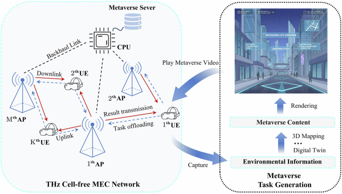
Terahertz Cell-Free Networks Harness AI to Power the Metaverse
New research introduces a novel network architecture combining Terahertz communication, cell-free MIMO, and AI-driven mobile edge computing to meet the extreme demands of the Metaverse.

Caterpillar Leverages Decades of Automation Experience for Broad AI Deployment
Caterpillar, a veteran in industrial automation, is applying its extensive experience from automating mining operations to the wider deployment of artificial intelligence across its products and internal processes.

Humanoid Robots: Evolution Beyond Expectation
The future of humanoid robots is unfolding in ways few predicted, moving past science fiction ideals to find practical applications in diverse sectors.