Predicting Cancer Genetics from Standard Biopsy Slides Using Computer Vision
Deep learning models are extracting actionable genetic alterations from standard H&E pathology slides, speeding up oncology decisions without consuming scarce tissue.
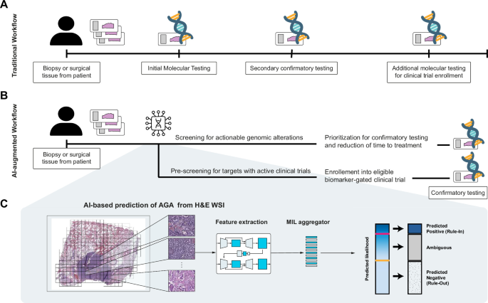
Deep learning models are increasingly able to detect critical cancer mutations directly from standard microscope slides, offering instant molecular insights without consuming scarce tissue or waiting weeks for sequencing.
The Bottleneck in Precision Oncology
Over the past two decades, precision oncology has rewritten the treatment playbook for solid tumors. In advanced non-small cell lung cancer, identifying actionable genomic alterations such as mutations in EGFR, ALK, or KRAS allows oncologists to bypass broad-spectrum chemotherapy in favor of targeted inhibitors. These targeted therapies can dramatically improve patient survival. Patients with EGFR mutations, for example, often experience progression-free survival exceeding two years when treated with third-generation tyrosine kinase inhibitors, compared to barely five months on standard platinum-based chemotherapy.
Yet the clinical promise of precision oncology frequently clashes with physical and logistical reality. Matching a patient to the right drug requires comprehensive genomic profiling via next-generation sequencing. To run these tests, laboratories need adequate tumor tissue. In practice, modern biopsies are often performed with fine needles or small core instruments, yielding minuscule tissue volumes.
Pathologists must divide this limited sample across multiple diagnostic tasks, including initial cancer confirmation, subtyping, and immunohistochemistry for protein targets. By the time genomic sequencing is ordered, the tissue is often exhausted. Furthermore, standard genomic assays typically take two to four weeks to return results. For patients experiencing rapid clinical deterioration, waiting a month for sequencing data is simply not an option. Studies of community oncology clinics reveal that up to 40 percent of patients with advanced lung cancer never receive recommended genomic testing before starting first-line therapy.
Reading Mutations from Routine Slides
To bypass this logjam, researchers have turned to artificial intelligence. Instead of dissolving tissue to extract and sequence DNA, computer vision models analyze the digital scans of standard histology slides.
Every diagnostic pathology workflow in the world begins with hematoxylin and eosin staining. This century-old technique dyes cell nuclei a deep purple-blue and cytoplasmic structures pink. While human pathologists use these routine slides to assess tumor architecture and grade, the human eye cannot reliably deduce specific underlying genetic mutations from visual appearance alone.
Deep learning systems, however, operate at a scale of pattern recognition that exceeds human perception. By ingesting whole-slide images digitized at high resolution, typically yielding images containing several billion pixels, convolutional neural networks and vision transformers can detect subtle morphological signatures associated with specific driver mutations. Genetic alterations alter cellular metabolism, nuclear geometry, stromal density, and spatial relationships between tumor cells and the microenvironment. Deep learning models map these microscopic nuances to the presence of specific alterations, such as EGFR or KRAS in lung adenocarcinoma, and FGFR in urothelial bladder carcinomas.
Because hematoxylin and eosin slides are generated for virtually every patient as part of the initial diagnosis, using AI to screen them requires no extra tissue cuts. The process is completely non-destructive, preserving the physical biopsy for confirmatory tests or orthogonal molecular assays.
Prospective Trials and Clinical Triaging
Until recently, the vast majority of digital pathology studies were retrospective, trained and tested on curated historical archives like The Cancer Genome Atlas. While these experiments proved the basic concept, retrospective datasets often mask real-world flaws, such as variable sample prep, inconsistent staining, and diverse patient demographics.
The field has now crossed into prospective clinical validation. Studies like the EAGLE and ANNAR clinical trials have demonstrated that pathology foundation models and fine-tuned deep learning pipelines can reliably identify genetic alterations across diverse, international patient cohorts.
Crucially, clinical advocates are not proposing that AI replace molecular sequencing outright. Current algorithms maintain high negative predictive value, meaning they are exceptionally good at ruling out specific alterations, but their positive predictive value can vary depending on mutation prevalence. As a result, the primary utility of these systems lies in clinical triaging.
When a biopsy is digitized, an algorithm can screen the image within minutes. If the model detects a strong probability of a rare or highly actionable mutation, the laboratory can immediately fast-track the sample for targeted confirmatory assays or liquid biopsies, skipping intermediate administrative delays. Conversely, in low-resource settings where access to comprehensive genomic profiling is scarce or financially prohibitive, an algorithmic screen can identify the select subset of patients most likely to benefit from expensive off-site genomic testing.
Technical Hurdles in Digital Pathology
Despite remarkable progress, transitioning image-based mutation prediction into routine clinical environments requires clearing significant technical barriers.
Histology slides are physical objects subject to tremendous operational variation. Different laboratories use different tissue processors, microtome slice thicknesses, staining reagents, and slide scanners. A digital image produced by an Aperio scanner looks subtly different in color balance and focus compared to one produced by a Hamamatsu or Roche system. Early models often suffered from site-specific bias, latching onto scanning artifacts or staining variations rather than true biological signals.
Addressing this issue has required the development of robust pathology foundation models trained on hundreds of thousands of diverse whole-slide images using self-supervised learning. These models learn generalizable representations of tissue biology that remain stable across scanner brands and laboratory protocols.
Computational overhead is another major factor. A single whole-slide image can exceed two gigabytes in size. Running multi-scale transformer architectures across dozens of gigapixel slides per day requires dedicated local compute or high-bandwidth uplinks to centralized processing centers. Pathology departments must integrate secure image management systems with laboratory information systems, ensuring that inference results flow directly to the reviewing pathologist without disrupting daily reporting.
As outlined in an analysis published in Nature, the shift from exploratory retrospective models to prospective clinical integration marks a pivotal phase for computational pathology. Realizing the full potential of these tools depends on establishing standardized regulatory pathways, clear performance thresholds, and seamless hospital network integration.
Why It Matters
The emergence of image-based genetic prediction illustrates how machine learning can extract hidden computational signals from existing physical infrastructure. Standard optical pathology has remained conceptually unchanged for generations, but digitizing the workflow transforms simple diagnostic glass into dense datasets.
For enterprise technology and infrastructure operators, this transition highlights the growing need for specialized edge compute and high-capacity local networking within healthcare facilities. Whole-slide scanners generate terabytes of uncompressed visual data weekly. Transmitting, storing, and running low-latency inference on these massive files demands robust data pipelines, dependable on-premises storage arrays, and optimized acceleration hardware deployed directly inside hospital data centers.
At the field level, the operational reliability of medical imaging hardware becomes paramount. Biomedical technicians and IT specialists are no longer just maintaining standalone microscopes or office networks. They are managing high-throughput imaging systems whose calibration, optical alignment, and data output directly inform artificial intelligence models that guide life-or-death oncology treatments.
More from Trends
RSS
Inside China's Coordinated Campaign to Build an AI-Native Generation
With mandatory curricula, entrance exam integration, and rural AI tutors, China is executing a massive state push to achieve universal AI literacy across 200 million students by 2030.
Securing Generative Biology: Inside the Defence-in-Depth Push for Biological AI
As AI models advance from generating protein structures to viable viral genomes, biosecurity researchers outline a multi-layered defence across data, models, and DNA synthesis.

Offshoring the Compute Boom: Washington Looks to Southeast Asia for Data Centers
Facing domestic grid bottlenecks and community backlash, US officials are promoting offshore AI infrastructure and chip manufacturing in the Philippines.