Mapping Cellular Recruitment: How Interpretable AI Decodes Spatial Biology
A deep learning framework called SPACER reads spatial transcriptomics data to reveal how cells recruit one another, offering clear rules for cancer immunity and therapy.
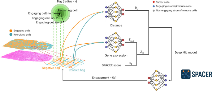
An interpretable neural network called SPACER decodes how cells recruit one another inside human tissue, transforming spatial transcriptomics into explainable biological rules.
Modern medicine has made rapid progress in identifying the genetic signatures of individual cells. Yet knowing what a cell expresses in isolation does not explain how it behaves within a crowded living tissue. In tumours, inflamed organs, and developing tissues, biological function depends entirely on location and neighbourhood interactions. Cells produce chemical gradients, alter physical barriers, and actively recruit other cell populations to their immediate vicinity.
Mapping these recruitment dynamics has remained a stubborn computational bottleneck. Standard computational tools can easily measure gene expression across thousands of cells, but they struggle to systematically determine why certain immune cells migrate toward specific cellular targets while avoiding others. Deep learning models can often predict clinical outcomes from raw spatial data, yet their internal logic is frequently opaque, functioning as black boxes that offer little actionable biology to researchers.
A new framework called SPACER tackles this limitation directly. By pairing spatial coordinates with gene expression data through an interpretable neural architecture, the system reveals the explicit operational rules that govern how target cells draw in cellular partners across tissue landscapes.
The Geometry of Cellular Microenvironments
Tissues are not random mixtures of cells. They are tightly orchestrated ecosystems where physical distance dictates chemical influence. Spatially resolved transcriptomics has emerged over recent years as a dominant method for capturing this complexity, allowing scientists to measure thousands of RNA transcripts while preserving the exact two-dimensional or three-dimensional coordinates of each cell.
Despite the power of this measurement technology, translating millions of data points into biological mechanisms requires structural assumptions. If an immune cell sits next to a malignant cell, researchers need to determine whether the immune cell was deliberately summoned, trapped by extracellular matrix, or merely passing through. Standard statistical correlations often mistake co-occurrence for deliberate recruitment.
To bridge this gap, computational biologists designed SPACER to model cell-cell engagement explicitly. The framework analyses tissue specimens by treating individual cells as pairs: a recruiting cell, such as a tumour cell, and an engaging cell, such as an immune cell or stromal component. By continuously evaluating the physical separation between these cells alongside their simultaneous gene expression profiles, the system calculates targeted scores that predict whether an active recruitment event has taken place.
Inside the Architecture of SPACER
Rather than feeding raw spatial maps into a dense convolutional or transformer network, SPACER relies on multiple instance learning. In this setup, training data are grouped into bags of cellular pairs. A positive bag contains cells that successfully form dense, interacting clusters, while a negative bag represents spatial configurations where recruitment failed to materialize.
For each pair in a bag, the model computes an interaction metric that accounts for two core variables: Euclidean distance and reciprocal molecular expression. If two cells are physically distant, their likelihood of active engagement drops sharply. Conversely, if two cells are close and express complementary molecular machinery, their pair score climbs.
The real breakthrough lies in the network's interpretability. Instead of obscuring its calculations behind deep, unreadable layers of artificial neurons, SPACER forces its internal representations through linear bottlenecks and explicit attention weights. Researchers can interrogate the trained weights to see precisely which genes, receptors, and environmental cues drove the prediction.
This design removes the guesswork typically associated with deep learning in spatial biology. Pathologists and researchers do not simply receive a binary prediction of cellular engagement. They receive an itemised ledger showing which biological pathways were necessary to recruit the interacting cell to that exact coordinates.
Decoding Tumour Defences and Therapy Complications
When deployed across complex clinical tissue datasets, SPACER immediately surfaced concrete biological rules that govern human disease. In multiple cancer types, the model illuminated the precise mechanisms that regulate how T cells enter the tumour microenvironment, a critical variable in determining whether a patient responds to immunotherapy.
The network revealed that T cell infiltration relies heavily on intact local antigen presentation machinery within the tumour cells themselves. When tumour cells downregulate these presentation markers, spatial recruitment drops, allowing the cancer to create cold microenvironments shielded from immune surveillance. Concurrently, the model pinpointed tumour mucins, heavily glycosylated proteins on the cell surface, as physical and biochemical deterrents that alter cellular recruitment rules and prevent immune penetration.
Beyond cancer infiltration, the system provided new insight into severe clinical side effects caused by cancer drugs. Immune checkpoint inhibitors can trigger fatal inflammatory conditions, most notably myocarditis, an intense inflammation of heart muscle tissue. By evaluating spatial transcriptomics data from animal models of severe checkpoint inhibitor-induced myocarditis, SPACER identified CD4+ T cells as primary effectors driving the cellular recruitment cascade in heart tissue.
This finding helps clarify a contentious debate in cardiology and oncology regarding which immune subsets spearhead tissue destruction in the heart during treatment complications. The research, published in Nature Biomedical Engineering, validated these computational predictions against advanced spatial receptor profiling methods, demonstrating that interpretable AI can generate testable, biologically accurate hypotheses from noisy clinical samples.
The Shift Toward Interpretable Scientific Systems
The development of SPACER reflects a broader, necessary shift within artificial intelligence for the life sciences. For several years, machine learning models in biology prioritised raw benchmark accuracy over transparency. In clinical settings and pharmaceutical pipelines, however, high accuracy without interpretability carries limited practical utility. A model that predicts patient deterioration or drug resistance without explaining the underlying mechanism cannot easily guide drug discovery or clinical trial design.
Interpretable architectures invert this dynamic. By constraining neural networks to conform to physical and biological principles, such as spatial decay over distance and known molecular binding principles, researchers turn AI from an arbitrary scoring engine into an analytical microscope. The model extracts reproducible biological logic that wet-lab scientists can test in animal models and clinical assays.
This methodological shift is also lowering computational overhead. Massive, unconstrained transformer models require enormous clusters and extensive compute time to process multi-gigabyte spatial imaging files. Constrained, multiple instance learning architectures like SPACER focus compute cycles strictly on the relevant local interactions, reducing training overhead while producing outputs that biological researchers can read and verify directly.
Why It Matters
For technical leaders, infrastructure specialists, and data engineers, the principles behind SPACER extend far beyond oncology and molecular biology.
Modern industrial facilities, telecom operations, and distributed data centres generate massive streams of spatial telemetry. Server racks dissipate heat across physical distances; microclimate sensors log temperature variations; automated hardware nodes communicate across local topologies. Diagnosing anomalies in these environments involves the same fundamental challenge seen in tissue biology: understanding how localized physical proximity and functional status interact to cause cascading system events.
As edge devices and high-throughput imaging systems are deployed closer to operating rooms, field laboratories, and clinical facilities, data pipelines must process high-resolution spatial datasets locally. Deploying massive, uninterpretable models to the edge is rarely feasible due to power, compute, and regulatory constraints. Architectures that bake physical geometry and explainable rules directly into the network design offer a blueprint for lighter, highly efficient edge analytics.
Whether tracing immune cell movement inside a malignant lesion or pinpointing thermal and signal degradation across a dense compute cluster, the future of diagnostic AI belongs to systems that can explain why an interaction took place, not just that it happened.
More from Trends
RSS
Auditing Medical Bias: Why Foundation Models Struggle with Clinical Fairness
A comprehensive review reveals how foundation models inherit and amplify healthcare disparities, exposing critical gaps in clinical testing and safety benchmarks.
Dendritic AI: How Brain Architecture Unlocked a 50-Million-Token Context Window
By replicating the non-linear processing of biological dendrites, researchers built a foundation model capable of processing 50 million tokens with lower compute.

Inside China's Relentless Drive for Scientific and Technological Primacy
China's state-backed push into fundamental research, semiconductors, and cost-efficient AI is systematically reshaping global technology competition.
